Fourier-Transform Infrared Spectrometer (FTIR)
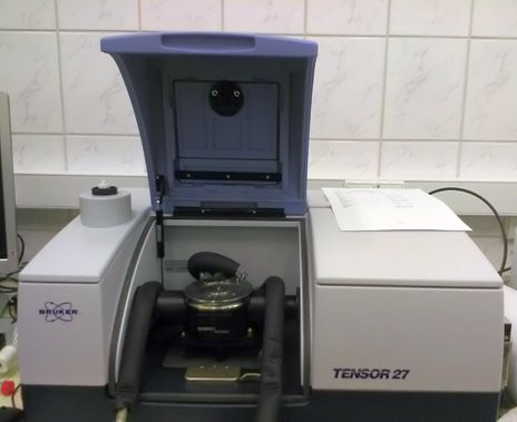
TENSOR 27 with AquaSpec and Bio-ATRII cells (Bruker)
Room 612
Contact: K. Skowronek
Access: collaboration/contracted research, trained users
General description
Spectrometer is dedicated to FTIR analysis of biological (aqueous) samples using two dedicated measurement cells: AquaSpec – transmittance cell with optical pathlength of ~ 7 µm and BioATRII, Attenuated Total Reflection cell. Measurements are performed in precisely regulated temperature 4 – 98 °C. Dedicated OPUS 7.0 software is used for data collection and analysis. Automatic measurements at temperatures ramps are possible.
Applications
- Protein secondary structure analysis. By analysis of amide I band (1600 -1800 cm-1) shape in FTIR spectrum, that corresponds to C=O stretching vibration within peptide bonds it is possible to measure secondary structure content of protein in a sample.
- Protein stability analysis. Spectra are recorded in increasing temperature and therefore changes in protein secondary structure induced by heating can be assessed.
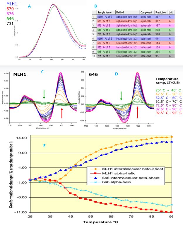
Analysis of single amino acid substitutions in MLH1 protein. A. Comparison of amide-I spectrum. B. Differences in secondary structures content estimated from FTIR spectra. C and D Differential spectra upon thermal denaturation. E. changes in secondary structures content induced by thermal denaturation (K. Poleszak)
Sample requirements
20 µl and ~50 µl of sample is required for BioATRII and for AquaSpec respectively. The protein concentration should be in 1 – 20 mg/ml range. Exactly matched buffer (preferably buffer after dialysis, filtrate or fractions preceding peak if sample is obtained directly form chromatography) is absolutely necessary for proper measurement of the background spectrum.
Further reading
- Bruker Application Note AN # 404 (https://www.bruker.com/fileadmin/user_upload/8-PDF-Docs/OpticalSpectrospcopy/FT-IR/CONFOCHECK/AN/AN404_Study_protein_conformation_EN.pdf)
- Fabian H and Mäntele HW. Infrared Spectroscopy of Proteins. doi: 10.1002/0470027320.s8201
- https://www.chem.uwec.edu/chem455_s05/pages/Manuals/FTIR_of_proteins.pdf
- Carbonaro M, Nucara A. Secondary structure of food proteins by Fourier transform spectroscopy in the mid-infrared region. Amino Acids. (2010) 38(3):679-90. doi: 10.1007/s00726-009-0274-3. PMID: 19350368.
- Kong J, Yu S. Fourier transform infrared spectroscopic analysis of protein secondary structures. Acta Biochim Biophys Sin (Shanghai). (2007) 39(8):549-59. PMID: 17687489.
1.